McMurray Test: Purpose, Procedure, and Clinical Interpretation
In breve
- McMurray’s test screens for suspected medial or lateral meniscal tears of the knee.
- With the patient supine, the clinician fully flexes the knee, rotates the tibia, applies valgus or varus stress, and extends the knee. A painful palpable or audible click supports a positive McMurray sign.
- A 2025 arthroscopy-validated study reported 60.9% sensitivity and 61.5% specificity for medial tears. For lateral tears, sensitivity was 55.6% and specificity was 96.3%.
- Clinicians should interpret McMurray’s test alongside the history, joint line tenderness, Thessaly test, and Apley’s compression test. A negative result does not reliably exclude a tear, and a positive result does not establish tear location or severity.
What the McMurray test screens for
McMurray’s test screens for suspected medial or lateral meniscal tears in the knee. The test provides a provocative finding rather than a standalone diagnosis because its diagnostic accuracy varies by compartment, population, and technique. A positive McMurray sign cannot reliably identify the exact tear pattern, size, or involved meniscal zone without other examination findings or imaging.
The maneuver loads meniscal tissue by combining tibial rotation with knee flexion and extension. As the knee extends, rotation and compartment-specific stress may catch displaced meniscal tissue between the femur and tibia, reproducing pain or a palpable or audible click. Clinical descriptions of meniscal lesions report medial tears more often than lateral tears and identify the posterior horn as the most frequently affected zone.
Clinicians should interpret McMurray’s test as one part of a meniscal assessment. A recent arthroscopy-validated study found modest and compartment-dependent accuracy, which supports pairing the maneuver with history, joint line tenderness, and other meniscal tests.
Performing the McMurray test step by step
-
Position the patient supine. Ask the patient to relax the tested leg and report any familiar pain, catching, or clicking during the maneuver. Stand beside the affected knee.
-
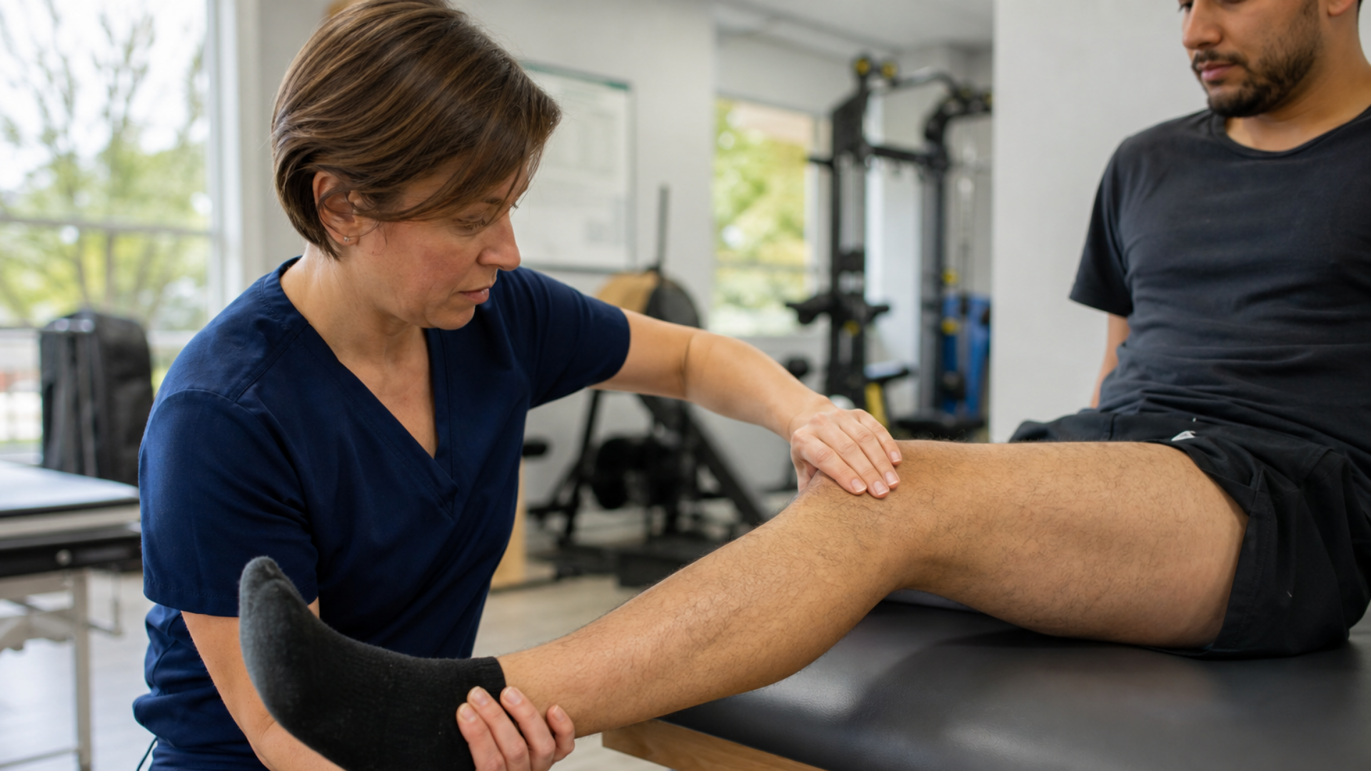
Place your hands to control and monitor the knee. Hold the heel with one hand so you can control knee flexion, extension, and tibial rotation. Place the other hand across the knee, with the thumb along the medial joint line and the fingers along the lateral joint line, so you can apply force and palpate movement.
-
Begin in maximal comfortable knee flexion. Avoid forcing flexion when pain, swelling, or restricted motion limits the available range. Establish whether symptoms are already present before adding rotation.
-
Stress the lateral meniscus. Internally rotate the tibia, apply a varus force at the knee, and slowly extend the knee through the available range. Palpate the lateral joint line and ask the patient to identify the location and character of any reproduced symptoms.
-
Stress the medial meniscus. Return the knee to flexion, externally rotate the tibia, and apply a valgus force while slowly extending the knee. Palpate the medial joint line and note any pain, click, pop, or catching. These rotation and loading directions follow the commonly described McMurray test technique.
-
Repeat only as needed to clarify the finding. Maintain controlled rotation and steady movement rather than applying abrupt force. Stop if the maneuver produces excessive pain or guarding that prevents meaningful assessment.
-
Document the full response. Record the tested side, available range, tibial rotation direction, applied valgus or varus force, symptom location, pain reproduction, and any palpable or audible click. Note whether guarding or restricted motion affected test quality.
Modified McMurray procedures may change the loading, movement range, or repetition pattern. Name the exact variant in the chart, and do not apply diagnostic-accuracy estimates from the standard maneuver to a modified version without evidence that evaluates that version.
Interpreting a positive finding
A positive McMurray sign requires a palpable or audible click or pop that reproduces the patient’s familiar joint-line pain during the maneuver. The examiner should record the knee position and movement phase at which the response occurs. Published descriptions sometimes count either pain or clicking as positive, so clinicians should document the observed components rather than rely on the word “positive” alone. The McMurray test description reflects this variation in criteria.
Pain without a click or pop provides a less specific result because joint irritation, osteoarthritis, or other knee pathology may produce pain during loaded rotation. Restricted motion without the characteristic response may reflect guarding, effusion, or baseline loss of range. A painless click does not meet the combined criterion used here.
The rotation direction used when the response occurs can suggest medial or lateral meniscal involvement according to the maneuver described above. However, McMurray’s test cannot confirm the tear pattern, exact location, size, or severity.
A chart note could read, “McMurray’s test reproduced familiar medial joint-line pain with a palpable click during extension under the medial-meniscus maneuver. No comparable response occurred with lateral-meniscus stress.”
Sensitivity, specificity, and what the evidence actually supports
A 2025 prospective study found modest and uneven diagnostic accuracy when researchers compared McMurray’s test with arthroscopy in 152 adults aged 20 to 40. For medial tears, sensitivity reached 60.9% and specificity reached 61.5%. For lateral tears, sensitivity reached 55.6%, while specificity reached 96.3%. The low sensitivity means a negative McMurray sign cannot reliably exclude either tear type. The high lateral specificity suggests that a positive result may carry more diagnostic weight for lateral pathology, but the study population limits generalization.
Historical findings vary too widely to support one universal accuracy figure. Hing and colleagues reported sensitivity ranges of 16% to 88% for medial tears and 25% to 79% for lateral tears across classic and modified McMurray maneuvers. Gupta and colleagues reported 50% sensitivity and 79% specificity for medial tears, while Akseki and colleagues reported 82% overall accuracy for lateral tears and 66% for medial tears.
Some apparently stronger results describe a full clinical examination rather than McMurray’s test alone. Ercin and colleagues reported 93% accuracy for an experienced surgeon’s combined clinical evaluation. Antinolfi and colleagues similarly reported 91% sensitivity, 87% specificity, and 90% accuracy for clinical examination in patients with suspected medial tears. Those figures should not be assigned to McMurray’s test as an isolated maneuver.
Study populations, examiner technique, test variants, and positive-test definitions all contribute to the spread in reported accuracy. A non-weight-bearing maneuver may also fail to reproduce symptoms that occur under load. Clinicians should therefore interpret sensitivity and specificity within the population and method used rather than applying one published number to every patient.
Joint line tenderness has stronger support as an isolated examination finding. A 2003 evidence-based guideline cited in the 2025 study identified joint line tenderness, rather than McMurray’s test, as the only standalone clinical test with reliable diagnostic value. McMurray’s test contributes more useful information when clinicians combine it with joint line tenderness and other meniscal findings.
Building a cluster: McMurray, Thessaly, Apley's, and joint line tenderness
Use a testing cluster to assess whether symptoms remain consistent across palpation, non-weight-bearing rotation, and weight-bearing rotation. Concordant findings can increase or decrease clinical suspicion, while disagreement may identify pain, guarding, or restricted motion that limits one maneuver.
-
Begin with the history and joint line tenderness. Ask about the injury mechanism, catching or locking, and effusion. With the knee flexed to 90 degrees, palpate the medial and lateral joint lines separately and compare them with the unaffected knee.
-
Perform the McMurray maneuver described above. Record the compartment, rotation direction, pain response, and any palpable or audible click rather than documenting only a positive or negative result.
-
Add Apley's test with the patient prone and the knee flexed to 90 degrees. Compress the tibia into the femur while rotating it, then repeat the rotation with distraction. Pain that increases during compression and decreases during distraction supports a meniscal source more than a ligamentous source, although Apley's test cannot confirm a tear by itself. The Apley's test mechanics differ from McMurray because the examiner compares axial compression with distraction.
-
Use the Thessaly test when weight bearing and rotational movement are appropriate. The patient stands on the test leg and rotates the body over a fixed foot at approximately 5 degrees and then 20 degrees of knee flexion. Joint line discomfort, catching, or locking constitutes a positive response. Thessaly applies rotation under axial load, which may reproduce symptoms that non-weight-bearing maneuvers do not. The standard Thessaly test procedure uses the unaffected leg first for familiarization.
Published evidence supports interpreting McMurray alongside other examination findings because its standalone accuracy varies substantially across populations and methods. However, pooled accuracy figures for these individual tests do not establish a validated combined likelihood ratio for this specific cluster. Treat concordance as clinical evidence within the broader examination, not as a separate diagnostic rule.
From findings to next steps: imaging, referral, or a conservative trial
Use the full presentation rather than a fixed McMurray threshold to choose the next step. A negative result cannot reliably exclude a tear because reported sensitivity remains modest. A positive result also carries different weight across populations. In one arthroscopy-validated study, sports-related injuries produced stronger diagnostic values than other mechanisms, while accuracy varied by age group. The study included adults aged 20 to 40, so those subgroup findings may not transfer to older patients.
MRI or referral becomes more reasonable when several findings support meniscal injury, symptoms persist, or imaging would change management. Discordance also matters. For example, a convincing history and joint line tenderness may justify further investigation despite a negative McMurray sign. MRI remains the most accurate noninvasive imaging option, while arthroscopy remains the reference standard for confirmation in research and surgical assessment according to an arthroscopy-validated diagnostic study.
A conservative rehabilitation trial may be reasonable when the examination does not indicate a need for prompt specialist assessment and current function permits nonoperative management. A positive test does not automatically place the patient on a surgical pathway. In moderate-to-severe knee osteoarthritis, arthroscopy has shown no significant benefit over optimized nonoperative therapy. You should therefore interpret a suspected meniscal lesion within the patient’s age, injury mechanism, osteoarthritis burden, functional limits, and response to rehabilitation.
From clinical findings to program design
Once clinical reasoning supports a conservative rehabilitation plan, Physitrack can help you deliver and monitor the prescribed exercises. The home exercise program builder lets you select content from more than 18,000 exercises and adjust the program to the patient’s current capacity, goals, and response.
PhysiApp delivers the program to the patient and records exercise completion at the session level. Patients can log completed sets and repetitions, along with pain and difficulty ratings. You can review those reports between visits and modify the program when your reassessment supports a change.
As with the prior Neer Test article in this special-test series, assessment findings should guide what happens next rather than dictate a standard program. Physitrack organizes prescription, delivery, and follow-up. It does not interpret the McMurray sign, establish a diagnosis, or replace the clinician’s reasoning.
Domande frequenti
How does McMurray’s test differ from Apley’s and Thessaly?
McMurray’s test applies tibial rotation while the clinician moves the supine knee through flexion and extension. Apley’s compares compression with distraction in prone, while Thessaly uses rotation over a weight-bearing leg. Each maneuver stresses the meniscus differently, so clinicians may use them together.
What does a false-positive McMurray sign mean?
A false positive occurs when the maneuver reproduces pain or a click despite no confirmed meniscal tear. Another painful knee structure, nonspecific clicking, or osteoarthritic change may contribute, but the test cannot identify the cause by itself. History, joint-line tenderness, other tests, and imaging when indicated help clarify the finding.
Can clinicians use McMurray’s test after surgery or with effusion and limited range of motion?
Postoperative restrictions, effusion, pain, or limited motion may prevent the knee movement required for a valid test. Clinicians should follow surgical precautions and avoid forcing flexion or rotation. An incomplete maneuver should be documented as limited rather than negative.
How reliable is McMurray’s test between examiners?
Inter-examiner reliability depends on consistent hand placement, force, movement, and interpretation of pain or clicking. Published accuracy varies substantially with technique and study population, which limits confidence in a universal reliability estimate. Clinicians should document the response precisely and interpret it within a broader examination rather than treating the McMurray sign as definitive.
Key takeaway
McMurray’s test provides one screening input for suspected meniscal injury. Arthroscopy-validated evidence shows modest and variable accuracy, so a positive McMurray sign cannot confirm a tear and a negative result cannot exclude one. Clinicians should interpret the finding alongside the history, joint line tenderness, other meniscal tests, and imaging when the broader presentation warrants it.

