Blood Flow Restriction Training: A Clinician's Guide to Safe Implementation
En resumen
- Blood flow restriction training pairs low-load exercise with a proximal cuff that limits arterial inflow and blocks venous outflow.
- Restricted flow accelerates metabolite buildup and muscle fatigue, which recruits higher-threshold motor units and stimulates muscle protein synthesis at loads around 20% to 30% of one-repetition maximum, with some literature supporting a broader 20% to 40% range.
- Clinical evidence most strongly supports BFR after ACL reconstruction and knee surgery, particularly when quadriceps weakness persists and heavy loading remains unsuitable.
- Clinicians must measure limb occlusion pressure, use individualized cuff pressures, and screen every patient for contraindications before treatment. Fixed pressures and systolic blood pressure proxies cannot account for differences in limb size, cuff design, or vascular response.
What blood flow restriction training is and why it works
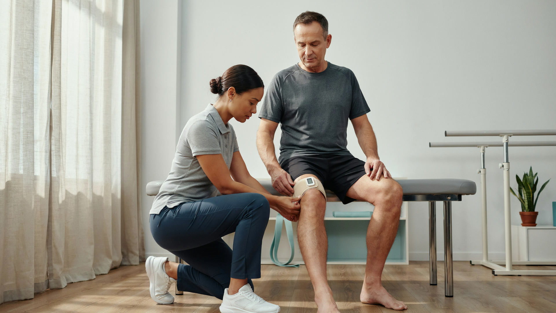
Blood flow restriction training combines low-load exercise with a pneumatic cuff placed near the top of an arm or leg. The cuff reduces arterial inflow and restricts venous return without fully stopping circulation. Clinicians commonly prescribe resistance at 20 to 30 percent of one-repetition maximum, compared with the 60 to 80 percent often used for conventional strength training.
Restricted blood flow makes a light set metabolically demanding. Reduced oxygen availability and slower metabolite clearance accelerate local fatigue, so the nervous system recruits additional motor units to maintain force. Type II fibers therefore contribute at loads that would ordinarily rely more heavily on fatigue-resistant fibers. A recent physiology review describes hypoxia and metabolite accumulation as major drivers of this early recruitment pattern and the resulting anabolic response (Frontiers in Physiology).
Cellular signaling adds to the training stimulus. Fluid accumulation within muscle fibers produces cell swelling, while the combined metabolic stress and mechanical tension activate pathways associated with protein synthesis. Research has implicated increased satellite cell activity, activation of the mTOR pathway, and reduced myostatin expression. Hormonal changes also occur, but current evidence does not establish any single hormone as the main cause of adaptation.
Low-load BFR generally produces greater hypertrophy and strength gains than the same load without occlusion. Findings summarized in a clinical review suggest that hypertrophy can approach the results of conventional high-load training, while maximal strength gains may remain greater with heavy resistance. The same review reports improved quadriceps size and strength when clinicians added low-load BFR during the first weeks after ACL reconstruction, with some studies also finding better knee-specific patient-reported outcomes and reductions in pain or effusion (clinical review).
BFR therefore gives physical therapists a way to create a substantial muscular stimulus when healing tissue, joint irritability, pain, or general load intolerance limits external resistance. Accurate cuff dosing and contraindication screening remain necessary because excessive pressure or effort changes both the stimulus and the risk profile.
Determining limb occlusion pressure and setting cuff pressure
Systolic blood pressure should not determine blood flow restriction cuff pressure. Patients with the same systolic blood pressure can have different limb occlusion pressures, so an SBP formula may underdose one patient and fully occlude another. Current guidance instead recommends individualized dosing at 40% to 80% of limb occlusion pressure.
Limb occlusion pressure, or LOP, is the minimum cuff pressure required to stop arterial blood flow beneath the cuff. For manual measurement, the clinician places the cuff proximally and monitors a distal arterial pulse by palpation or Doppler while gradually inflating the cuff. LOP occurs when the pulse disappears during inflation, although a descending method can identify the pressure at which the pulse returns during deflation. Manual accuracy depends on operator skill, equipment sensitivity, and consistent technique.
Automated cuffs measure LOP by detecting arterial pulsations with a sensor placed on a finger or toe while the cuff inflates in controlled steps. Clinicians should determine LOP using the same cuff and placement planned for exercise because cuff width, shape, bladder length, and material affect the required pressure. A fixed value applied to every patient, such as a flat 300 mmHg practice standard, does not represent the same degree of restriction across different patients or devices.
Equipment selection should account for pressure control during movement. In one comparison at a target of 80% LOP, the four tested devices required average pressures ranging from 135 to 191 mmHg. The devices also differed in how consistently they stayed near the prescribed target during exercise. Clinicians should favor cuffs that measure individualized LOP and maintain the selected pressure under changing muscle tension rather than assuming the displayed setting remains stable.
BFR protocol table by clinical population
Post-ACLR has the clearest postoperative evidence, including studies of low-load BFR during early rehabilitation. Reviewed ACLR protocols commonly use 20% to 30% of 1RM and the 30/15/15/15 repetition scheme. Protocols for non-ACL postoperative quadriceps atrophy and older adults draw more heavily on general BFR guidance, so clinicians should treat those ranges as starting points rather than fixed prescriptions.
Current guidance supports individualized pressures between 40% and 80% of measured LOP. Your surgical protocol, tissue-healing constraints, symptom response, and exercise tolerance should determine the final dose. When a patient cannot complete the planned repetitions with acceptable symptoms, reduce pressure or load before increasing rest or changing the exercise.
Where the evidence is strongest: post-ACL reconstruction and post-surgical quad atrophy
Post-ACL reconstruction offers the clearest clinical rationale for early BFR because quadriceps loss begins quickly while heavy resistance remains poorly tolerated. Patients may lose 20% to 33% of quadriceps muscle volume between the injury and three weeks after surgery. Disuse contributes to that loss, while surgery-related neural inhibition reduces voluntary quadriceps activation and limits the loading available through conventional exercise.
Clinical studies suggest that BFR can help preserve tissue and strength during this low-load phase. A synthesis of five postoperative studies, citing Aspetar's ACL guidelines, found that adding low-load BFR may improve quadriceps and hamstring strength and limit early disuse atrophy. The small Jack et al. study followed 32 patients with bone-patellar tendon-bone grafts and began BFR during postoperative week two. Patients who completed BFR alongside standard rehabilitation preserved more bone mass on DXA than the control group. The study supports BFR as an adjunct, but its sample size and graft-specific population limit broader conclusions.
Clinicians should treat evidence for general post-surgical quadriceps atrophy as less direct. ACL reconstruction studies provide the strongest support, while protocols for other knee surgeries often extend those findings to patients facing similar restrictions on heavy loading. Surgical precautions, graft or tissue healing, weight-bearing status, and irritability should still determine exercise selection and progression.
Adolescents may need a slower pressure progression than adults. One adolescent protocol started patients as young as 12 at 80% limb occlusion pressure about one week after surgery. Participants reported minor effects such as itching, but dropout was relatively high. Investigators linked poor tolerance partly to starting at the full target pressure, so clinicians should consider increasing pressure gradually across the first few sessions rather than applying 80% immediately.
Older adults and load-intolerant patients
Blood flow restriction training can provide a lower-load strength option for older adults and patients who cannot tolerate conventional heavy resistance. Exercises performed at 20 to 30% of one-repetition maximum reduce joint and tissue loading while the cuff helps produce the metabolic stress and motor unit recruitment described above. Published guidance supports a broader working range of 20 to 40% of one-repetition maximum.
Lower loads may suit patients whose pain, frailty, arthritis, or medical status limits heavy lifting. BFR can help bridge the period between basic activation work and progressive resistance training, but clinicians should still progress toward conventional loading when the patient can tolerate it and the treatment plan calls for it.
Evidence for older and load-intolerant populations remains thinner than the evidence for ACL reconstruction. Clinicians should avoid assuming that ACLR findings apply equally to every older adult or chronic condition. Individual screening, LOP-based pressure, symptom monitoring, and reassessment should guide use in these patients.
Contraindications and screening before starting BFR
A pre-BFR screen should occur before pressure selection or exercise testing. Clinicians should document vascular history, pregnancy status, relevant medications, resting blood pressure, skin condition, and prior surgery that may affect circulation or lymphatic drainage.
The contraindications below reflect commonly cited clinical screening categories rather than a single peer-reviewed consensus list. Treat them as a starting framework for a clinic policy, and confirm against current professional guidelines and physician input for complex cases.
Absolute contraindications
For a conservative clinic policy, do not apply BFR when the patient has any of the following conditions.
- Active or previous deep vein thrombosis, pulmonary embolism, or a known clotting disorder
- Pregnancy
- Peripheral vascular disease or clinically significant poor circulation
- Active cancer or a tumor in the treatment limb
- An open wound, active infection, or fracture in the treatment area
A medical review may change how a specific case is classified, but clinicians should not proceed until the relevant risk has been assessed and documented.
Relative contraindications
The following conditions warrant medical clearance and an individualized risk assessment before BFR begins.
- Uncontrolled hypertension
- Anticoagulant or antiplatelet medication that affects clotting
- Varicose veins
- Previous lymph node removal
- Diabetes with vascular complications
- Sickle cell anemia
Clinicians should also consider the patient’s current cardiovascular status, sensation, skin integrity, and ability to report symptoms accurately. Clearance does not replace individualized limb occlusion pressure measurement or monitoring during exercise.
Available adverse-event claims require careful interpretation. A single physical therapy clinic page is also the source for the contraindications list above, and that page reports that properly screened and dosed BFR produced few adverse events without linking its claims to traceable peer-reviewed studies. Clinicians should not treat either the contraindications categories or the adverse-event statements as confirmed against a systematic review, and should still apply independent clinical judgment and physician consultation for complex cases.
Temporary muscle swelling, mild lightheadedness, or brief tingling may occur around cuff release. Persistent numbness, unusual discoloration, disproportionate pain, shortness of breath, or other unexpected symptoms should prompt immediate cuff removal and clinical assessment. Each clinic should pair its screening form with an escalation policy and staff training before offering BFR.
Is BFR safe and does it work: clinician FAQ
Is BFR training safe?
BFR appears safe for appropriately screened patients when clinicians individualize pressure and control exercise dosage. Reported temporary effects include bruising, muscle soreness, and paresthesia, while serious adverse events have been associated with inappropriate use or excessive loading in the clinical literature. Clinicians should apply the contraindications screening described above before every initial prescription and reassess when health status changes.
Does BFR training work?
Low-load BFR can improve muscle strength and size when pain, surgical precautions, or joint irritability limit heavy resistance exercise. Evidence supports its use after ACL reconstruction and for postoperative weakness, although outcomes depend on suitable cuff pressure, exercise dosage, and progression. BFR should complement progressive resistance training rather than replace heavier loading once the patient can tolerate it.
What equipment does BFR require?
Clinical BFR requires a cuff that fits the limb, a way to measure limb occlusion pressure, and equipment that maintains the prescribed pressure during exercise. Cuff width affects the pressure needed for occlusion, so clinicians should use the manufacturer’s sizing guidance and document the cuff used. Pneumatic systems can measure and reproduce pressure more precisely than elastic wraps, which do not provide a dependable percentage of limb occlusion pressure.
How often can patients perform BFR?
Most strengthening protocols use two or three sessions per week. Short early postoperative protocols may use more frequent sessions, but clinicians should adjust frequency for soreness, swelling, wound status, and exercise tolerance. Each session should follow the prescribed restriction time and allow reperfusion between exercises when the protocol requires it.
Who should supervise BFR training?
A clinician trained in BFR screening, limb occlusion pressure measurement, cuff placement, and adverse-response management should supervise initial sessions. Selected patients may later perform prescribed BFR exercises outside the clinic after demonstrating correct cuff use and understanding when to stop. The supervising physical therapist should review symptoms, adherence, and loading before progressing pressure or resistance.
Building BFR into a tracked home exercise program
Home BFR should begin only after you confirm that the patient can position the cuff, use the prescribed device, and follow the stop criteria independently. Build the home exercise program with the prescribed cuff pressure as a percentage of limb occlusion pressure, exercise load, sets, repetitions, rest periods, and weekly frequency. Include clear instructions for symptoms that require the patient to stop and contact the clinic.
PhysiApp records exercise completion, sets and repetitions, and patient-reported pain and difficulty for each session. Physitrack sends those session-level records to the clinician dashboard, so you can compare prescribed dosage with what the patient completed between visits. A missed final set or rising difficulty rating gives you more useful context than a login record alone.
Physitrack carries your clinical protocol between appointments, but it does not select cuff pressure or confirm that the patient applied it correctly. You should continue to reassess limb occlusion pressure, cuff technique, symptoms, and exercise tolerance in person. Use the reported data to guide progression, reinforce technique, or return BFR sessions to supervised care when needed.
The takeaway for clinics adding BFR to their caseload
Clinics can adopt BFR safely when they treat individualized LOP dosing and contraindication screening as required clinical controls. A sensible rollout starts with trained clinicians, a small caseload, and cuffs that support reliable LOP measurement.
Clinic directors should also define documentation and escalation procedures before expanding access. Track prescribed pressure, completed sets, symptoms, and adherence within the patient’s broader home exercise program. Those records help clinicians adjust dosage between visits and identify cases that require reassessment.

